By: Dr. Robert L. Bard Edited by: L. Goetze, Ed.D / C. R. DeWitt / G. Davi
IMAGING HEART HEALTH
Carotid ultrasound is also crucial for detecting early atherosclerosis, a key driver of heart disease. By evaluating plaque buildup and arterial thickness, it identifies cardiovascular risks before major events occur. Since heart disease risk rises post-menopause, early vascular screening allows for timely preventive strategies.
Incorporating ultrasound into routine assessments enables early intervention, guiding lifestyle and medical management to reduce heart disease risks. Given that cardiovascular disease is the leading cause of death in women, proactive imaging supports a healthier transition into menopause. Incorporating ultrasound into routine assessments enables early intervention, guiding lifestyle and medical management to reduce heart disease risks. Given that cardiovascular disease is the leading cause of death in women, proactive imaging supports a healthier transition into menopause.
SLIDE 1: DETECTING SILENT MYOCARDIAL AND CARDIOVASCULAR DISEASE
Many years ago, the Framingham study, conducted for life insurance purposes, identified carotid intima-media thickness (CIMT) as an indicator of decreased life expectancy. Since that study, which is now over 20 years old, further research has shown that the lining of the carotid artery can thicken up to one centimeter. CIMT is now recognized as a key health marker and a measure of longevity.
Advanced imaging techniques, such as super-selective B-flow ultrasound, allow for precise measurement of CIMT. In this case, the ultrasound scan shows a CIMT of 1.2 millimeters. The adjacent image highlights the underlying cause: inflammatory vessels associated with arteritis. These inflamed vessels contribute to arterial wall thickening, even in the absence of significant arterial blockage. A microvascular imaging scan of the same carotid artery further demonstrates the contrast between normal and thickened arterial walls.
Although the artery may not be obstructed, individuals with a CIMT exceeding 1.0 millimeters face an increased risk of stroke. The rightmost image presents an innovative technique for assessing the hardness of arterial plaques. Soft plaques are more prone to embolization and can travel to the brain, whereas calcified plaques are generally more stable. Shear wave ultrasound technology enables early detection of arterial stiffness, providing insight into vascular health before overt disease develops. In addition to measuring arterial inflammation and fibrosis, this method helps evaluate the extent of arterial wall scarring.
During menopause, most myocardial and cardiovascular events occur silently. Women experiencing heart attacks are often misdiagnosed with chest pain, pruritus, or musculoskeletal discomfort. However, a simple one-minute carotid artery scan can help detect silent myocardial and cardiovascular disease, making it a valuable tool for identifying potential serious conditions in asymptomatic menopausal patients.
One incidental benefit of scanning the thyroid for tumors or inflammatory disease is the ability to assess nearby structures, including lymph nodes—where cancer may spread—and the carotid artery, which supplies blood to the brain. In this case, the arrow indicates plaque in the carotid artery, measuring 1.3 millimeters in thickness. Since the normal threshold is 1.0 millimeter, this finding suggests an early potential cardiovascular issue. This discovery is relevant to both thyroid and heart disease. Additionally, the scan reveals an incidental thyroid cyst and an abnormal, non-homogeneous thyroid.
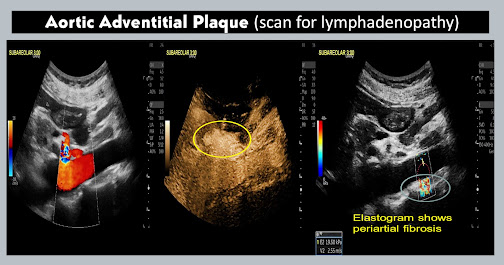
When scanning for aortic aneurysms or aortic adenopathy to assess whether breast cancer has spread to the lymph node chain, we typically observe the aorta as a midline vessel. In this case, during scanning, we detected abnormal echoes in the peri-aortic region. To further investigate, a Doppler blood flow scan was performed, which ruled out an aneurysm. Subsequently, an aortic scan with blood flow analysis was conducted to assess abnormal arterial activity, revealing that the thickened area had no discernible arterial inflammatory blood flow. However, the elastogram on the far right indicated abnormal fibrosis of the aortic wall, which could weaken the structure and potentially lead to rupture.
SLIDE 4: THYROID SCAN WITH POINT OF CARE ULTRASOUND
Portable high-resolution thyroid scanning is clinically available. In the labeled scan, the thyroid tissue appears milky white, while a dark area represents a cyst. We can confirm this is a cyst because, to its left, the carotid artery—filled with blood—also appears dark. A magnified scan (third image) reveals that this is not a simple cyst. The cyst wall contains internal micro-calcifications, and a follow-up study indicates inflammation. The presence of red arterial flow suggests irritation of the cyst wall. I could tag the middle image, which clearly shows the carotid artery.

SUPPLEMENTAL
The Many Faces of Heart Disease and Cardiovascular Disease
By: Lennard Goetze, Ed.D and Graciella Davi
Heart disease and cardiovascular disease (CVD) are umbrella terms encompassing a wide range of conditions that affect the heart and blood vessels. These conditions can lead to serious complications, including heart attacks, strokes, and even death. Understanding the various types of heart disease can help individuals recognize symptoms early, seek appropriate treatment, and take preventative measures to maintain heart health.
 1) CORONARY ARTERY DISEASE (CAD) is one of the most common types of heart
disease. It occurs when the coronary arteries, which supply oxygen-rich blood
to the heart, become narrowed or blocked due to plaque buildup
(atherosclerosis). This can lead to chest pain (angina), shortness of breath,
and heart attacks. Lifestyle changes, medications, and procedures such as
angioplasty or bypass surgery can help manage CAD.
2) HEART FAILURE, also known as congestive heart failure (CHF), occurs when
the heart cannot pump blood effectively to meet the body’s needs. It can result
from conditions such as CAD, high blood pressure, or cardiomyopathy. Symptoms
include fatigue, shortness of breath, and fluid retention. Treatment includes
medications, lifestyle changes, and in severe cases, devices like pacemakers or
heart transplants.
3) ARRHYTHMIAS are irregular heartbeats that can be too fast (tachycardia), too slow (bradycardia), or erratic. These abnormal rhythms can interfere with the heart’s ability to pump blood effectively. Causes range from heart disease and electrolyte imbalances to stress and medications. Treatments include medications, lifestyle modifications, and procedures such as ablation or the implantation of pacemakers.
4) HEART VALVE DISEASE occurs when one or more of the heart’s valves do not
function properly, leading to issues such as stenosis (narrowing of the valve)
or regurgitation (leakage of blood backward). Common symptoms include fatigue,
chest pain, and swelling in the legs. Treatment options vary from medication
management to surgical repair or replacement of the affected valve.
5) CARDIOMYOPATHY refers to diseases of the heart muscle that make it harder
for the heart to pump blood efficiently. Types include dilated, hypertrophic,
and restrictive cardiomyopathy. Symptoms often include breathlessness, swelling
in the legs, and irregular heartbeats. Treatment depends on the type but may
involve medications, lifestyle changes, and in severe cases, heart transplants.
6) CONGENITAL HEART DISEASE (CHD) refers to structural heart abnormalities present at birth. These defects can range from minor to severe and may involve holes in the heart, abnormal connections between blood vessels, or improperly formed valves. Some cases require surgical correction, while others may be managed with medications and ongoing monitoring.
8) PERIPHERAL ARTERY DISEASE (PAD) occurs when arteries outside the heart,
particularly in the legs, become narrowed due to atherosclerosis. This can lead
to pain, numbness, and even tissue damage in severe cases. PAD increases the
risk of heart attack and stroke. Treatment focuses on lifestyle changes,
medications, and procedures to restore blood flow.
9) AORTIC DISEASE affects the body’s main artery, the aorta. Conditions such
as aortic aneurysm (a bulging or weakened area in the aorta) or aortic
dissection (a tear in the artery’s wall) can be life-threatening. Symptoms
include severe chest or back pain, dizziness, and difficulty breathing.
Treatment ranges from monitoring to emergency surgical repair.
10) RHEUMATIC HEART DISEASE is caused by rheumatic fever, an inflammatory disease that can damage heart valves. It often develops from untreated or inadequately treated strep throat infections. Symptoms include breathlessness, fatigue, and irregular heartbeats. Prevention through early treatment of strep infections is crucial, and management includes medications and sometimes valve surgery.
11) STROKE occurs when blood flow to the brain is disrupted, either due to a
blocked artery (ischemic stroke) or a ruptured blood vessel (hemorrhagic
stroke). This can cause brain damage, disability, or death. Symptoms include
sudden weakness, confusion, trouble speaking, and loss of coordination.
Treatment involves clot-busting drugs, surgery, and rehabilitation.
12) PULMONARY EMBOLISM (PE) occurs when a blood clot, usually from a DVT, travels to the lungs, blocking blood flow. This can be life-threatening, causing sudden shortness of breath, chest pain, and rapid heart rate. Immediate treatment with anticoagulants, thrombolytics, or surgery is necessary to prevent fatal complications.
Conclusion
Heart disease and cardiovascular disease manifest in many forms, each with its own causes, symptoms, and treatment options. Early detection, lifestyle modifications, and appropriate medical intervention are key to managing these conditions and improving overall heart health. Regular check-ups, maintaining a balanced diet, engaging in physical activity, and managing stress can significantly reduce the risk of developing these life-threatening diseases.
RELATED ARTICLE
Understanding Thyroid Health: Key Insights on Hormones, Longevity, and Wellness
Thyroid health plays a critical role in nearly every physiological process of the body, influencing metabolism, brain function, heart health, and more. Yet, despite its significance, the nuances of thyroid function remain elusive for many. As we age, maintaining optimal thyroid function is an essential component of overall well-being, and understanding the balance of thyroid hormones can help prevent future health challenges. See Dr. Angela Mazza's full report on Thyroid Health @ this season's MenoNews.
For more information on this article, or to explore diagnostic imaging related to cardiovascular health, visit: www.menoscan.org or www.BardDiagnostics.com. You can also contact our editorial team at: 516-522-0777, or email us at: editor.prevention101@gmail.com
www.BARDDIAGNOSTICS.com
212.355.7017
© Copyright 2025- Bard Diagnostics LLC.& the AngioFoundation 501c3. All Rights Reserved.


Dr. Bard leads the charge! The information in this message highlights the critical relationship between menopause and cardiovascular health, really emphasizing how declining estrogen levels contribute to increased heart disease risk through negative changes in cholesterol levels, blood pressure, and arterial stiffness.
ReplyDeleteUltrasound can play a critical role in early detection of cardiovascular health changes, offering a non-invasive method for assessments. which can lead to preventive measures. This underscores the importance of proactive cardiovascular screening, especially for menopausal women, preventing misdiagnosis and shortening of quality of life.